Thanks to gravity and the aging process, a certain amount of spinal compression is natural as you get older. But for some people, the narrowing of the spinal canal can lead to pain and other symptoms. With more and more people suffering from back pain, it’s important to be aware of your spine and understand how to keep it healthy.
A healthy spine
The human spine should curve in an S-shape – an important design for our bipedal (two-legged) upright sitting or standing stance. It allows the weight distribution to shift as we move and holds our head and organs vertically up from the ground.
Our spines consist of 24 individual sections of bone (vertebrae) separated by facet joints, which contain rubbery discs of cartilage to keep the whole spinal column freely mobile (see the illustration on page 41, top image). This also creates space for the spinal canal, a cavity running through each of the vertebrae, which encloses the spinal cord, spinal nerves, ligaments, fat and blood vessels.
Spinal nerves exit the spinal canal through nerve root canals (intervertebral foramen) to branch out into your whole body. These canals are surrounded by bone and ligaments, and changes in the bone structure can narrow them and create compression or restriction of the spinal cord or nerves. Pain or numbness can occur when blood flow out to the limbs is restricted.
Spinal stenosis
One cause of narrowing of the spine is spinal stenosis – a degenerative condition where any combination of bone spurs, enlarged facet joints or bulging discs constrict the nerve root canals, causing compression and entrapment of the spinal nerves and even the spinal cord, called central stenosis (see page 41, second image from top). It can occur along any area of the spine: the neck (cervical), upper back (thoracic) or most commonly the lower back (lumbar).
Spinal stenosis is typically caused by changes in the spine related to osteoarthritis and often accompanies herniated discs (page 41, third image), where a portion of the inner soft center of the disc (the nucleus) pushes through the harder exterior (annulus). Symptoms of spinal stenosis will follow if the herniation compresses a nerve.
Since modern postural habits such as sitting for long periods of time are inherently compressive for the spine, some people with spinal stenosis may not be aware of its presence or experience any symptoms. As you can see from the bottommost diagram on page 41, modern postural patterns that deviate away from our ideal alignment can create compression in various forms, depending on individual tendencies.
Signs of spinal stenosis
Pain that is relieved by rest or bending forward, where the spine is flexed, and worsened by spine extension or bending backward, could signal spinal stenosis. Other signs are tingling, numbness and muscle weakness.
When stenosis affects the cervical spine (neck), symptoms are usually felt in the arms, and when in the lower back,most often in the legs. Symptoms can worsen over time, with loss of bladder and bowel control in the most severe cases involving the lower back.
The conventional medical view is that there’s no cure for spinal stenosis, and spinal ‘decompression’ surgery may be offered. Usually, this will involve a laminectomy, which removes the lamina – the posterior (back) part of a vertebra (spinal bone) that covers your spinal canal. This is most commonly performed in the lumbar area (the lower back).
The surgery essentially enlarges the spinal canal to relieve pressure on the spinal cord or nerves, but according to a 2017 review,1 it comes with a significantly greater risk of complications compared to physical therapy programs. Risks of surgery include recurrent or continuing symptoms, infection, blood clots, leakage of cerebrospinal fluid, nerve injury, paralysis and loss of vision.
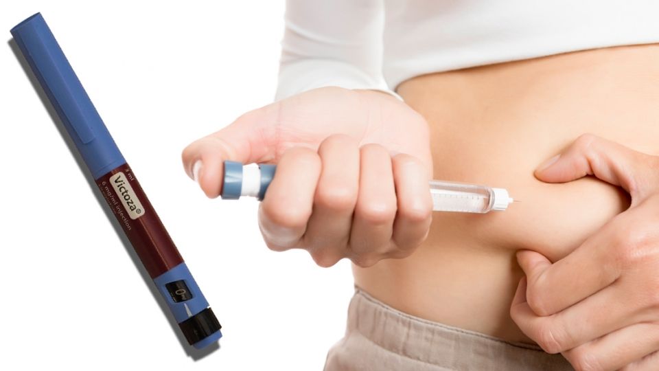
Patients are also routinely offered high-dose pain medication for the inflammatory effects that create chronic pain in the back and legs, as the nerves feeding down into the legs are impinged, although this pain can often be caused by muscle imbalance and not the stenosis per se (see WDDTY May 2019).
Physiotherapy may be advised to help increase the spinal range of motion and ensure that sufferers do not avoid movement, which they often do because of the pain. Further loss of range of motion can worsen symptoms.
The myofascia (muscle and connective tissue) in all areas of the body – including around the spine – need constant motion to bring in nutrients and oxygen, as well as remove waste products from throughoutthe body. This keeps tissues hydrated and less prone to adhesions (fibrous bands that form between tissues and organs) or hardening, which can add to inflammation and transmit pain to other areas too.
A reliance on pain medication can worsen the situation, as it not only reduces beneficial gut bacteria and encourages inflammation,2 but also masks the pain, which could otherwise inform us how best to move.
The importance of balance
Those with spinal stenosis have been historically recommended to only bend the spine forward, putting it into flexion. This was believed to relieve nerve pressure by increasing the diameter of the spinal canal. However, moving in one direction without the balance of the opposite restricts full motion of the body.
The exercise sequence described on the following pages includes some gentle back bending, because although deeper back arches can put too much pressure onto discs that may be herniated (closing the spinal canal further), gently pressing in that area can move nerves away from the spinal canal, offering them more space and helping to relieve compression.
Balance is important too. Encouraging healthy posture with natural spinal curves relies on balance between the front and back body, so becoming aware of both of those as you open the spine front and back can also help inform the way you move in daily life.
Ultimately, if you can feel uplift through the spine whenever possible, there is less nerve impingement. Strengthening the muscles that support the spine upward also creates the space that relieves compression.
Exercises for relieving spinal compression
The following exercises are mostly isometric in nature – where muscles contract but don’t actually move, i.e, they hold you in place with muscle fibers activated but with equal forces working against each other.
This creates a strengthening effect and, without the exertion of movement, also allows you to breathe fully and does not contribute to any stress response, which fires off protective, inflammatory reactions.
The most important guide within these exercises is to listen and respond to what feels right for your body at any given time. If you find that any posture aggravates your symptoms or sets off internal alarm bells, back off to a place where it feels you can soften and breathe with the physical sensations.
If there is no position that feels safe, stop doing the exercise. However, with each final position there is a gentle pulsing motion to help the myofascia become more pliable in that direction. You can explore this to help loosen tissues locked into patterns that exacerbate symptoms.
Some side-bending and twisting is included, as moving away from the source of pain can decrease pressure on nerve roots for those with lateral stenosis. So if the pain is on the right, a bend or twist to the left may make space on the right-hand side and relieve pressure
and pain there. Turning to the left would then need to be done very gently, with deep listening and feeling out the range of motion, which may differ greatly to the right.
The exercises are floor-based and can be a very helpful if done before a short or long walk to allow for the natural side-to-side motions of the spine, freeing up through the chest and shoulders. They can also be used to create space in the spine in preparation for other exercises.

Spinal fluidity
• From lying with feet at least hip-width apart, as you inhale, raise the pelvis into a bridge pose, lifting into the chest rather than the lower back. You don’t need to come up far – start halfway to gauge the sensations and feel the support of drawing up the belly. Your arms can come up and over the shoulders to reach above your head (as shown) or stay down by your hips as the pelvis lifts – each has different sensations in the back and spine, so see what suits you.
• On the exhale, lower the spine back down, vertebra by vertebra.
• Lift up and down, eventually holding the pose up for as long as you feel neither stress nor strain in the body or breath.
• Roll to your side to counter the pose with the gentle forward bend of a side-lying fetal position.

Exploring your spinal range of motion
• Sit in a’z-legs’position, with the left leg bent in and the right bent out in a wide seat and the left hand on the ground. Lift the right arm, bent so that the forearm is parallel to the ground. Inhale length in the spine and exhale to twist to the left, continuing this motion comfortably.
• Then with either the left elbow or hand on the ground, take the right arm up to where you can comfortably breathe easily as you lengthen the right side of the body and spine. This may be up by the right ear, reaching out at shoulder height or lower if need be.
• Come to the other side, respecting different needs on each side.

Spine suspended on all fours
• On all fours, begin moving into the shoulders and hips to tune in to how you feel around the spine. Gently ‘wag your tail’ from side to side for a little lateral motion.
• From a central position, with the inhale, arch your back just as much as you can easily open the chest without pain.
• With the exhale, draw your belly in to curve your back out and open the back body. Move between these two positions to alternately find space in the front and back of the spine.

Lengthening your spine
• From all fours, tuck the toes under and lift the hips. As you exhale, bring shoulders over wrists, coming right up onto the balls of the feet. Draw the chin into the chest, rounding the back and gathering the belly into the body.
• On the inhalation draw the thighs back toward downward-facing dog, opening the front body, so knees may be bent and heels high, coaxing out the pose with a rocking rhythm back and forth.
• If you settle into downward-facing dog pose, you can retain length in the spine and notice if tightness in the back of the legs or the top of the back limits that length. If so, keep the heels high so you can still retain openness in the chest.

Opening the front body
• Either from all fours or downward-facing dog, step your right foot forward so it is pointing straight ahead. From this lunge, bring your left hand onto your belly and right hand onto your lower back, feeling how you support uplift through the front and softening down the back.
• Taking the left hand to the outside of the right thigh, lift the left arm with elbow bent so the forearm follows the line of the collarbone. Then inhale into a twist and exhale to retract back again. Move to the other side.

Gentle resting spine flexion
Resting the back in a gentle forward bend allows you to integrate the previous movements and is a place to come back to if you need to relieve pressure at any time. A fetal position with feet on the floor or legs lengthened out (knees out to the sides) allows you to fully rest.

References
1 Int J Surg, 2017; 44: 329-38
2 Clin Microbiol Infect, 2016; 22: 178.e1-9
What do you think? Start a conversation over on the... WDDTY Community